
Diepoxy-treated bovine jugular vein conduit for pulmonary artery replacement
 N.R. Nichay
- I.Yu. Zhuravleva
- Yu.Yu. Kulyabin
- I.S. Zykov
- E.V. Boyarkin
- O.Yu. Malakhova
- E.V. Kuznetsova
- T.P. Timchenko
- Ya.L. Rusakova
- I.S. Murashov
- A.A. Dokuchaeva
- A.V. Bogachev-Prokophiev
Published 2022-12-29
Keywords
- Animals,
- Follow-Up Studies,
- Heart Valve Diseases,
- Jugular Veins,
- Right Ventricular Outflow Tract Reconstruction
How to Cite
Copyright (c) 2022 Nichay N.R., Zhuravleva I.Yu., Kulyabin Yu.Yu., Zykov I.S., Boyarkin E.V., Malakhova O.Yu., Kuznetsova E.V., Timchenko T.P., Rusakova Yu.L., Murashov I.S., Dokuchaeva A.A., Bogachev-Prokophiev A.V.

This work is licensed under a Creative Commons Attribution 4.0 International License.
Abstract
Objective: To evaluate the performance and short-term capacity of the diepoxy-treated bovine jugular vein conduit in large animals during 6-month follow-up.
Methods: Thirteen diepoxy-treated bovine jugular vein conduits were implanted into the pulmonary artery of young mini-pigs. During the follow-up, graft function was tested using transesophageal echocardiography. The animals were withdrawn at 6 months, and the explanted conduits were assessed histologically.
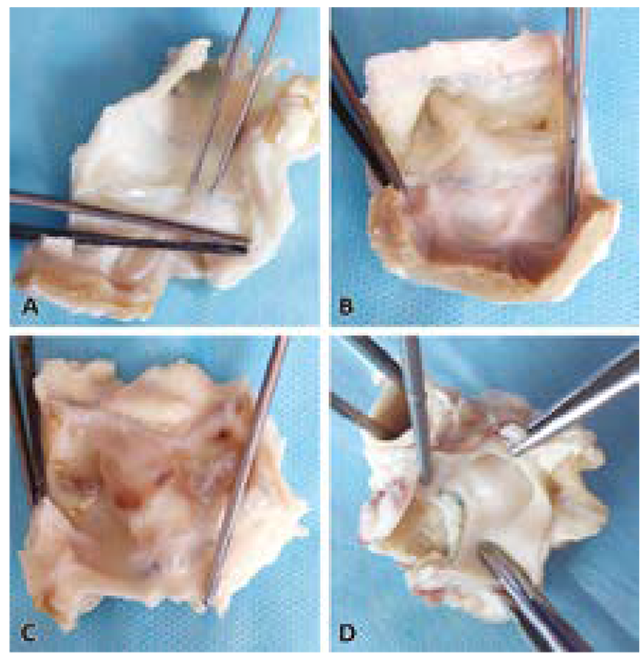
Results: All the conduits were successfully implanted without any surgical complications. All the animals survived throughout the follow-up. By the end of the follow-up period, the pressure gradient increased on five animals’ conduits including one case of mismatch between the conduit and the native pulmonary artery, two cases of distal stenosis, and two case of endocarditis. No significant increase in valve regurgitation or conduit thrombosis was observed during the follow-up. In conduits without dysfunction, the structure of the walls and leaflets was intact. A thin fibrous tissue covered the conduit inner wall with complete surface endothelialization. Neither signs of degeneration or calcification nor inflammatory cells were found in the conduit wall or leaflets. Neointima proliferation without calcium deposits was observed in two distally stenosed conduits. Inflammatory cells consisting of multinucleated macrophages, lymphocytes, and histiocytes were found in the adventitia. There were no inflammatory cells in the media or intima, and the leaflets showed no changes.
Conclusion: Diepoxy-treated bovine jugular vein demonstrated acceptable performance, good endothelialization, and low tendency to thrombosis and calcium accumulation in the wall and leaflets.
Received 31 October 2022. Revised 25 November 2022. Accepted 28 November 2022.
Funding: The study was supported by Russian Science Foundation (grant No. 22-25-20102).
Conflict of interest: The authors declare no conflict of interest.
Contribution of the authors
Conception and study design: N.R. Nichay, I.Yu. Zhuravleva, A.V. Bogachev-Prokophiev
Data collection and analysis: N.R. Nichay, Yu.Yu. Kulyabin, I.S. Zykov, E.V. Boyarkin, O.Yu. Malakhova, E.V. Kuznetsova, T.P. Timchenko, Ya.L. Rusakova, I.S. Murashov, A.A. Dokuchaeva
Statistical analysis: N.R. Nichay, I.Yu. Zhuravleva
Drafting the article: N.R. Nichay, I.Yu. Zhuravleva, Yu.Yu. Kulyabin, I.S. Zykov, E.V. Boyarkin, O.Yu. Malakhova, T.P. Timchenko, Ya.L. Rusakova, I.S. Murashov, A.A. Dokuchaeva
Critical revision of the article: N.R. Nichay, I.Yu. Zhuravleva, Yu.Yu. Kulyabin, A.A. Dokuchaeva, A.V. Bogachev-Prokophiev
Final approval of the version to be published: N.R. Nichay, I.Yu. Zhuravleva, Yu.Yu. Kulyabin, I.S. Zykov, E.V. Boyarkin, O.Yu. Malakhova, E.V. Kuznetsova, T.P. Timchenko, Ya.L. Rusakova, I.S. Murashov, A.A. Dokuchaeva, A.V. Bogachev-Prokophiev
References
- Yong M.S., Yim D., d'Udekem Y., Brizard C.P., Robertson T., Galati J.C., Konstantinov I.E. Medium-term outcomes of bovine jugular vein graft and homograft conduits in children. ANZ J Surg. 2015;85(5):381-385. PMID: 25708132. https://doi.org/10.1111/ans.13018
- Sandica E., Boethig D., Blanz U., Goerg R., Haas N.A., Laser K.T., Kececioglu D., Bertram H., Sarikouch S., Westhoff-Bleck M., Beerbaum P., Horke A. Bovine jugular veins versus homografts in the pulmonary position: an analysis across two centers and 711 patients-conventional comparisons and time status graphs as a new approach. Thorac Cardiovasc Surg. 2016;64(1):25-35. PMID: 26322831. https://doi.org/10.1055/s-0035-1554962
- Falchetti A., Demanet H., Dessy H., Melot C., Pierrakos C., Wauthy P. Contegra versus pulmonary homograft for right ventricular outflow tract reconstruction in newborns. Cardiol Young. 2019;29(4):505-510. PMID: 30942148. https://doi.org/10.1017/S1047951119000143
- Urso S., Rega F., Meuris B., Gewillig M., Eyskens B., Daenen W., Heying R., Meyns B. The Contegra conduit in the right ventricular outflow tract is an independent risk factor for graft replacement. Eur J Cardiothorac Surg. 2011;40(3):603-609. PMID: 21339072. https://doi.org/10.1016/j.ejcts.2010.11.081
- Patel P.M., Tan C., Srivastava N., Herrmann J.L., Rodefeld M.D., Turrentine M.W., Brown J.W. Bovine jugular vein conduit: a mid- to long-term institutional review. World J Pediatr Congenit Heart Surg. 2018;9(5):489-495. PMID: 30157735. https://doi.org/10.1177/2150135118779356
- Meyns B., Van Garsse L., Boshoff D., Eyskens B., Mertens L., Gewillig M., Fieuws S., Verbeken E., Daenen W. The Contegra conduit in the right ventricular outflow tract induces supravalvular stenosis. J Thorac Cardiovasc Surg. 2004;128(6):834-840. PMID: 15573067. https://doi.org/10.1016/j.jtcvs.2004.08.015
- Gist K.M., Mitchell M.B., Jaggers J., Campbell D.N., Yu J.A., Landeck B.F. 2nd. Assessment of the relationship between Contegra conduit size and early valvar insufficiency. Ann Thorac Surg. 2012;93(3):856-861. PMID: 22300627. https://doi.org/10.1016/j.athoracsur.2011.10.057
- Mery C.M., Guzmán-Pruneda F.A., De León L.E., Zhang W., Terwelp M.D., Bocchini C.E., Adachi I., Heinle J.S., McKenzie E.D., Fraser C.D. Jr. Risk factors for development of endocarditis and reintervention in patients undergoing right ventricle to pulmonary artery valved conduit placement. J Thorac Cardiovasc Surg. 2016;151(2):432-439. PMID: 26670191. https://doi.org/10.1016/j.jtcvs.2015.10.069
- Gröning M., Tahri N.B., Søndergaard L., Helvind M., Ersbøll M.K., Ørbæk Andersen H. Infective endocarditis in right ventricular outflow tract conduits: a register-based comparison of homografts, Contegra grafts and Melody transcatheter valves. Eur J Cardiothorac Surg. 2019;56(1):87-93. PMID: 30698682. https://doi.org/10.1093/ejcts/ezy478
- Imamura E., Sawatani O., Koyanagi H., Noishiki Y., Miyata T. Epoxy compounds as a new cross-linking agent for porcine aortic leaflets: subcutaneous implant studies in rats. J Card Surg. 1989;4(1):50-57. PMID: 2519982. https://doi.org/10.1111/j.1540-8191.1989.tb00256.x
- Umashankar P.R., Mohanan P.V., Kumari T.V. Glutaraldehyde treatment elicits toxic response compared to decellularization in bovine pericardium. Toxicol Int. 2012;19(1):51-58. PMID: 22736904; PMCID: PMC3339246. https://doi.org/10.4103/0971-6580.94513
- Grabenwöger M., Sider J., Fitzal F., Zelenka C., Windberger U., Grimm M., Moritz A., Böck P., Wolner E. Impact of glutaraldehyde on calcification of pericardial bioprosthetic heart valve material. Ann Thorac Surg. 1996;62(3):772-777. PMID: 8784007.
- Eybl E., Griesmacher A., Grimm M., Wolner E. Toxic effects of aldehydes released from fixed pericardium on bovine aortic endothelial cells. J Biomed Mater Res. 1989;23(11):1355-1365. PMID: 2558116. https://doi.org/10.1002/jbm.820231111
- Zhuravleva I.Y., Nichay N.R., Kulyabin Y.Y., Timchenko T.P., Korobeinikov A.A., Polienko Y.F., Shatskaya S.S., Kuznetsova E.V., Voitov A.V., Bogachev-Prokophiev A.V., Karaskov A.M. In search of the best xenogeneic material for a paediatric conduit: an experimental study. Interact Cardiovasc Thorac Surg. 2018;26(5):738-744. PMID: 29346675. https://doi.org/10.1093/icvts/ivx445
- Xi T., Ma J., Tian W., Lei X., Long S., Xi B. Prevention of tissue calcification on bioprosthetic heart valve by using epoxy compounds: a study of calcification tests in vitro and in vivo. J Biomed Mater Res. 1992;26(9):1241-1251. PMID: 1429769. https://doi.org/10.1002/jbm.820260913
- Chang Y., Sung H., Chiu Y., Lu J. Assessment of an epoxy-fixed pericardial patch with or without ionically bound heparin in a canine model. Int J Artif Organs. 1997;20(6):332-340. PMID: 9259210.
- Nishi C., Nakajima N., Ikada Y. In vitro evaluation of cytotoxicity of diepoxy compounds used for biomaterial modification. J Biomed Mater Res. 1995;29(7):829-834. PMID: 7593021. https://doi.org/10.1002/jbm.820290707
- Zhuravleva I.Yu., Karpova E.V., Dokuchaeva A.A., Kuznetsova E.V., Vladimirov S.V., Ksenofontov A.L., Nichay N.R. Bovine jugular vein conduit: What affects its elastomechanical properties and thermostability? J Biomed Mater Res A. 2022;110(2):394-408. PMID: 34390309. https://doi.org/10.1002/jbm.a.37296
- Nichay N.R., Zhuravleva I.Y., Kulyabin Y.Y., Zubritskiy A.V., Voitov A.V., Soynov I.A., Gorbatykh A.V., Bogachev-Prokophiev A.V., Karaskov A.M. Diepoxy- versus glutaraldehyde-treated xenografts: outcomes of right ventricular outflow tract reconstruction in children. World J Pediatr Congenit Heart Surg. 2020;11(1):56-64. PMID: 31835985. https://doi.org/10.1177/2150135119885900
- Никитин С.В., Князев С.П., Шатохин К.С. Миниатюрные свиньи ИЦиГ — модельный объект для изучения формообразовательного процесса. Вавиловский журнал генетики и селекции. 2014;18(2):279-293. Nikitin S.V., Knyazev S.P., Shatokhin K.S. Miniature pigs of ICG as a model object for morphogenetic research. Vavilov Journal of Genetics and Breeding. 2014;18(2):279-293. (In Russ.).
- Itoh T., Kawabe M., Nagase T., Endo K., Miyoshi M., Miyahara K. Body surface area measurement in laboratory miniature pigs using a computed tomography scanner. J Toxicol Sci. 2016;41(5):637-644. PMID: 27665773. https://doi.org/10.2131/jts.41.637
- Stout K.K., Daniels C.J., Aboulhosn J.A., Bozkurt B., Broberg C.S., Colman J.M., Crumb S.R., Dearani J.A., Fuller S., Gurvitz M., Khairy P., Landzberg M.J., Saidi A., Valente A.M., Van Hare G.F. 2018 AHA/ACC guideline for the management of adults with congenital heart disease: executive summary: a report of the American College of Cardiology/American Heart Association Task Force on Clinical Practice Guidelines. J Am Coll Cardiol. 2019;73(12):1494-1563. PMID: 30121240. https://doi.org/10.1016/j.jacc.2018.08.1028
- Lemson M.S., Tordoir J.H., Daemen M.J., Kitslaar P.J. Intimal hyperplasia in vascular grafts. Eur J Vasc Endovasc Surg. 2000;19(4):336-350. PMID: 10801366. https://doi.org/10.1053/ejvs.1999.1040
- Schoenhoff F.S., Loup O., Gahl B., Banz Y., Pavlovic M., Pfammatter J.-P., Carrel T.P., Kadner A. The Contegra bovine jugular vein graft versus the Shelhigh pulmonic porcine graft for reconstruction of the right ventricular outflow tract: a comparative study. J Thorac Cardiovasc Surg. 2011;141(3):654-661. PMID: 21255796. https://doi.org/10.1016/j.jtcvs.2010.06.068
- Ichikawa Y., Noishiki Y., Kosuge T., Yamamoto K., Kondo J., Matsumoto A. Use of a bovine jugular vein graft with natural valve for right ventricular outflow tract reconstruction: a one-year animal study. J Thorac Cardiovasc Surg. 1997;114(2):224-233. PMID: 9270640. https://doi.org/10.1016/S0022-5223(97)70149-1
- Flameng W., Hermans H., Verbeken E., Meuris B. A randomized assessment of an advanced tissue preservation technology in the juvenile sheep model. J Thorac Cardiovasc Surg. 2015;149(1):340-345. PMID: 25439467. https://doi.org/10.1016/j.jtcvs.2014.09.062
- Berdajs D., Mosbahi S., Vos J., Charbonnier D., Hullin R., von Segesser L.K. Fluid dynamics simulation of right ventricular outflow tract oversizing. Interact Cardiovasc Thorac Surg. 2015;21(2):176-182. PMID: 25912476. https://doi.org/10.1093/icvts/ivv108
- Wiebe D., Megerman J., L'Italien G.J., Abbott W.M. Glutaraldehyde release from vascular prostheses of biologic origin. Surgery. 1988;104(1):26-33. PMID: 3133800.
- Grimm M., Eybl E., Grabenwöger M., Griesmacher A., Losert U., Böck P., Müller M.M., Wolner E. Biocompatibility of aldehyde-fixed bovine pericardium. An in vitro and in vivo approach toward improvement of bioprosthetic heart valves. J Thorac Cardiovasc Surg. 1991;102(2):195-201. PMID: 1678026.
- Noishiki Y., Hata C., Tu R., Shen S.H., Lin D., Sung H.W., Witzel T., Wang E., Thyagarajan K., Tomizawa Y. Development and evaluation of a pliable biological valved conduit. Part I: Preparation, biochemical properties, and histological findings. Int J Artif Organs. 1993;16(4):192-198. PMID: 8325696.
- Patel S.D., Waltham M., Wadoodi A., Burnand K.G., Smith A. The role of endothelial cells and their progenitors in intimal hyperplasia. Ther Adv Cardiovasc Dis. 2010;4(2):129-141. PMID: 20200200. https://doi.org/10.1177/1753944710362903
- Scavo V.A. Jr, Turrentine M.W., Aufiero T.X., Sharp T.G., Brown J.W. Valved bovine jugular venous conduits for right ventricular to pulmonary artery reconstruction. ASAIO J. 1999;45(5):482-487. PMID: 10503630. https://doi.org/10.1097/00002480-199909000-00022
- Sung H.W., Shen S.H., Tu R., Lin D., Hata C., Noishiki Y., Tomizawa Y., Quijano R.C. Comparison of the cross-linking characteristics of porcine heart valves fixed with glutaraldehyde or epoxy compounds. ASAIO J. 1993;39(3):M532-M536. PMID: 8268592.
- Köstering H., Mast W.P., Kaethner T., Nebendahl K., Holtz W.H. Blood coagulation studies in domestic pigs (Hanover breed) and minipigs (Goettingen breed). Lab Anim. 1983;17(4):346-349. PMID: 6678359. https://doi.org/10.1258/002367783781062262
- Sondeen J.L., de Guzman R., Amy Polykratis I., Dale Prince M., Hernandez O., Cap A.P., Dubick M.A. Comparison between human and porcine thromboelastograph parameters in response to ex-vivo changes to platelets, plasma, and red blood cells. Blood Coagul Fibrinolysis. 2013;24(8):818-829. PMID: 24047887. https://doi.org/10.1097/MBC.0b013e3283646600
- Herijgers P., Ozaki S., Verbeken E., Van Lommel A., Meuris B., Lesaffre E., Daenen W., Flameng W. Valved jugular vein segments for right ventricular outflow tract reconstruction in young sheep. J Thorac Cardiovasc Surg. 2002;124(4):798-805. PMID: 12324739. https://doi.org/10.1067/mtc.2002.121043
- Attmann T., Quaden R., Freistedt A., König C., Cremer J., Lutter G. Percutaneous heart valve replacement: histology and calcification characteristics of biological valved stents in juvenile sheep. Cardiovasc Pathol. 2007;16(3):165-170. PMID: 17502246. https://doi.org/10.1016/j.carpath.2007.01.002
- Peivandi A.D., Seiler M., Mueller K.-M., Martens S., Malec E., Asfour B., Lueck S. Elastica degeneration and intimal hyperplasia lead to Contegra conduit failure. Eur J Cardiothorac Surg. 2019;56(6):1154-1161. PMID: 31280306. https://doi.org/10.1093/ejcts/ezz199